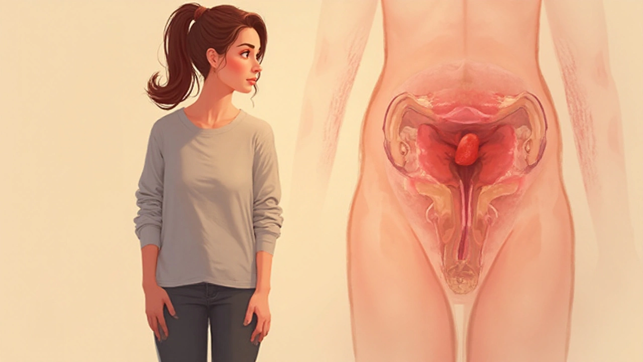
Endometrial hyperplasia is a benign proliferation of the cells lining the uterus (the endometrium), usually triggered by an imbalance of estrogen and progesterone. When this imbalance persists, the lining thickens beyond normal limits, setting the stage for bleeding problems and, in some cases, cancer.
Understanding the Endometrium
The endometrium is the inner mucosal layer of the uterus that sheds each month during menstruation. Its thickness naturally oscillates between 3mm (menstrual phase) and 14mm (mid‑secretory phase). Any sustained stimulus that pushes the endometrium to stay in the proliferative state leads to endometrial hyperplasia.
Hormonal Drivers Behind Overgrowth
Two hormones dominate endometrial behavior:
- Estrogen is a steroid hormone that stimulates endometrial cell division. Chronic estrogen excess-often seen in obesity, polycystic ovary syndrome (PCOS), or unopposed estrogen therapy-keeps the lining in a growth phase.
- Progesterone counteracts estrogen by promoting secretory transformation and limiting proliferation. Progesterone deficiency, whether due to anovulation or inadequate hormone replacement, removes this brake.
The combination of high estrogen and low progesterone creates the perfect storm for the endometrium to overgrow.
Types of Endometrial Hyperplasia
Clinicians distinguish three main categories based on architectural patterns and cytologic atypia. The most critical split is between typical (low‑risk) and atypical (high‑risk) forms.
| Feature | Typical Hyperplasia | Atypical Hyperplasia |
|---|---|---|
| Architectural pattern | Gland crowding without irregular shapes | Glandular irregularities, cribriform patterns |
| Cytologic atypia | Absent or mild | Marked nuclear enlargement, loss of polarity |
| Risk of progression to cancer | ~2% | ~30% over 5years |
| Management focus | Progestin therapy, surveillance | High‑dose progestins or hysterectomy |
From Thickening to Cancer
When atypical hyperplasia persists, genetic mutations accumulate-most commonly in PTEN and DNA mismatch‑repair pathways. These alterations can transform the hyperplastic tissue into endometrial carcinoma, the most common gynecologic cancer in developed nations. Early detection of atypia therefore cuts the pathway to invasive disease.
Diagnosing Overgrowth
Two tools dominate the workup:
- Transvaginal ultrasound measures endometrial thickness. In post‑menopausal women, a threshold of 5mm usually prompts further evaluation.
- Endometrial biopsy provides tissue for histologic grading, confirming typical or atypical forms.
Both tests are quick, outpatient procedures and together achieve >90% sensitivity for detecting significant hyperplasia.

Management Strategies
Therapeutic choices aim to restore hormonal balance and, when needed, remove abnormal tissue.
- Progestin therapy: Oral medroxyprogesterone acetate (10-20mg daily) or levonorgestrel‑releasing intra‑uterine system (IUS) delivers localized progesterone, reversing typical hyperplasia in 70‑80% of cases.
- Addressing underlying causes: Weight loss (5% body weight can cut estrogen production by 10%), treating PCOS with metformin, or stopping unopposed estrogen drugs such as tamoxifen in breast‑cancer survivors.
- Surgical options: For refractory atypical hyperplasia, hysterectomy offers definitive cure; fertility‑preserving high‑dose progestins are an alternative for younger women.
Related Conditions That Feed the Problem
Several health issues share the same estrogen‑dominant environment:
- Polycystic ovary syndrome (PCOS) often features chronic anovulation, leading to prolonged estrogen exposure.
- Obesity increases peripheral aromatization of androstenedione to estrone, raising circulating estrogen.
- Hormone replacement therapy (HRT) that includes estrogen without adequate progesterone can directly cause hyperplasia.
- Tamoxifen acts as an estrogen agonist in the uterus while blocking it in breast tissue, a double‑edged sword for cancer patients.
Screening women with these conditions for endometrial thickness is a cost‑effective way to catch hyperplasia early.
Practical Checklist for Patients and Clinicians
- Ask about menstrual irregularities, especially prolonged or heavy bleeding.
- Measure BMI; advise a target < 30kg/m² for estrogen‑related risk reduction.
- Review medication list for unopposed estrogen sources (HRT, tamoxifen, phytoestrogen supplements).
- Schedule a transvaginal ultrasound if post‑menopausal endometrial thickness > 5mm.
- Perform an endometrial biopsy when ultrasound or symptoms suggest hyperplasia.
- Start progestin therapy promptly for typical hyperplasia; reassess with repeat biopsy in 3-6 months.
- Consider hysterectomy for persistent atypical hyperplasia or when childbearing is complete.
Future Directions & Research Gaps
While the link between estrogen excess and hyperplasia is well‑established, gaps remain:
- Precise molecular signatures that predict which typical hyperplasia will evolve.
- Long‑term outcomes of levonorgestrel IUS in obese vs non‑obese populations.
- Non‑hormonal agents (e.g., metformin) as adjuncts to progestin therapy.
Ongoing trials are expected to refine risk‑stratification tools, making personalized treatment possible within the next few years.
Frequently Asked Questions
What symptoms suggest endometrial hyperplasia?
The most common sign is abnormal uterine bleeding-either heavy, prolonged, or spotting between periods. Some women notice pelvic pressure or a feeling of fullness, but many are asymptomatic and are diagnosed during routine screening.
How is a diagnosis confirmed?
First, a transvaginal ultrasound measures endometrial thickness. If the measurement exceeds the normal threshold (5mm post‑menopause) or bleeding patterns are abnormal, an office‑based endometrial biopsy is performed. The tissue sample is examined under a microscope and graded as typical or atypical hyperplasia.
Can lifestyle changes reverse hyperplasia?
Yes-weight loss of about 5% of body weight can lower estrogen production from fat, improving hormone balance. Regular aerobic exercise, a Mediterranean‑style diet, and smoking cessation also reduce the risk of progression.
Is the levonorgestrel IUS safe for all women?
The IUS is widely used and effective, but it’s contraindicated in women with active pelvic infection, unexplained vaginal bleeding, or uterine anomalies. A quick ultrasound helps rule out these issues before insertion.
When is surgery recommended?
Surgery (hysterectomy) is advised for persistent atypical hyperplasia that does not respond to high‑dose progestins, for women who have completed childbearing, or when an invasive cancer is suspected based on biopsy results.
How does tamoxifen affect the uterus?
Tamoxifen blocks estrogen receptors in breast tissue but mimics estrogen in the uterus. This partial agonist effect can stimulate the endometrium, increasing the risk of hyperplasia and, over time, carcinoma. Regular ultrasound monitoring is advised for long‑term users.
Richie Lasit
Wow, this is actually one of the clearest explanations I’ve seen on endometrial hyperplasia. I’ve been helping my sister through this and honestly, most docs just say ‘take progesterone’ and leave it at that. This breaks it down without dumbing it down. Huge props to the author.
arthur ball
Bro. I had this. Like, full on atypical. Took me 18 months to get someone to listen. They thought I was just ‘perimenopausal.’ Then I went full detective mode-tracked my meds, lost 12 lbs, got the IUS, and now my next biopsy’s clean. You’re not alone. And yes, weight loss isn’t ‘just a suggestion’-it’s medicine.
Justice Ward
This post reads like a love letter to the endometrium-respectful, detailed, and deeply human. I’m a nurse in OB/GYN and I’ve seen too many women dismissed because their bleeding was ‘just irregular.’ This kind of clarity? It saves lives. Seriously, someone should turn this into a patient handout.
maria norman
So let me get this straight-we’re treating a hormonal imbalance with synthetic hormones… while ignoring that the root cause might be environmental estrogens from plastics, soy, or even your damn shampoo? Interesting how the medical-industrial complex prefers a pill over a paradigm shift. 🤔
katerine rose
so like if you have pcos you’re basically doomed right like why even try
Jessie Bellen
They never tell you the real reason: Big Pharma wants you on tamoxifen, then on progestins, then on hysterectomies. The real cure? Stop estrogen mimickers. Look at your toothpaste. Your deodorant. Your takeout containers. This isn’t medicine-it’s profit-driven distraction.
lisa zebastian
Typical hyperplasia has a 2% cancer risk? That’s statistically negligible. The real risk is the iatrogenic trauma of unnecessary biopsies, progestin side effects (mood swings? fatigue? weight gain?), and the psychological terror of being labeled ‘pre-cancerous’ for a condition that often regresses spontaneously. This whole framework is fear-based medicine dressed in jargon. The data doesn’t support the panic.
Chris Rowe
lol u guys think this is complicated? in my village in nigeria we just drink bitter leaf juice and pray. no biopsies. no ius. no meds. just god and herbs. why u all so scared of nature?
Sushmita S
so like i had this and i just ate a lot of papaya and it went away?? idk man 🍉
Francis Pascoe
YOU’RE ALL MISSING THE POINT. The endometrium doesn’t ‘overgrow.’ It’s being attacked by electromagnetic frequencies from 5G towers, amplified by glyphosate in your food, and weaponized by the CDC to control female fertility. The biopsy? A trap. The IUS? A tracking device. They want you docile. Wake up.
Iris Schaper
my aunt had this and she just started doing yoga every day and now she’s fine. no meds. no surgery. just breathin. maybe we’re all overcomplicating this?
Selma Cey
If estrogen is the villain, then why is it essential for bone density, brain function, and cardiovascular health? Are we really going to treat a physiological response as a pathology? This feels like blaming the messenger. The real question: why is our society producing so much estrogenic stress? Not the endometrium’s fault.
Harrison Dearing
Y’all are losing your minds. 😭 I had atypical hyperplasia. I did the progestin. I lost weight. I got the IUS. I’m fine now. It’s not a conspiracy. It’s biology. And if you’re reading this and scared? Go see a doc. Not Reddit. Just go. 💪
Jasmine Kara
i just read this and i’m like… wait so i dont have to get a hysterectomy?? my dr said i did but now i’m not sure… i’m gonna ask again
bhuvanesh kankani
In India, we have traditionally used Ashoka bark and Shatavari to regulate hormonal balance. While modern medicine offers precision, these botanicals have been used for millennia with documented efficacy. Integrative approaches may offer gentler pathways for those seeking alternatives to synthetic hormones.
Richa Shukla
soo like tamoxifen is bad for uterus but good for breast?? so it’s like… giving cancer to one part to save another?? why tf do they even give this??
AnneMarie Carroll
Oh please. You all think this is just about hormones? It’s about control. The system wants women to be scared of their own bodies. That’s why they push biopsies and hysterectomies. They don’t want you to know you can heal naturally. They want you dependent. Wake up. This isn’t medicine. It’s maintenance.